
































by Stephane Frouel, Julie Castier & Maxime Hugonin, MiXscience, France
Aquaculture is a complex industry with multiple variables that strongly impact its performance.
Among these, sources of stress, disease outbreaks and occurrence of new pathogens such as parasites are a huge threat.
Enterocytozoon hepatopenaei (EHP) is a microsporidian parasite that was first characterised in the black tiger shrimp Penaeus monodon in 2009 in Thailand. EHP is restricted to the shrimp hepatopancreas (HP) and its occurrence was usually given scant attention because of the overwhelming focus on some other diseases, such as early mortality syndrome (EMS) and white faeces syndrome. However, although EHP does not appear to cause high mortality, it is associated with severe growth retardation and depressed feed conversion ratio (FCR) in shrimp, leading to economic losses for the farmers who may need to resort to an early harvest.
To date, no reliable and sustainable solutions have shown real efficacy on the reduction of impact of EHP in shrimp. The aim of the current research at miXscience (France), is to design a specific solution against EHP in shrimp aquaculture. Efficacy was demonstrated from the laboratory to the field, focusing on the reduction of germination of EHP spores and its associated effect on shrimp performance. Complementary studies have been performed to explore, by microscopy, the fine mechanisms of action of the solution and demonstrate how this new product disturbs the EHP spores.
A reminder: The EHP cycle in shrimp
The life cycle of this microsporidian parasite can be divided into three phases: the infective, the proliferative and the spore-forming phase [Figure 1].
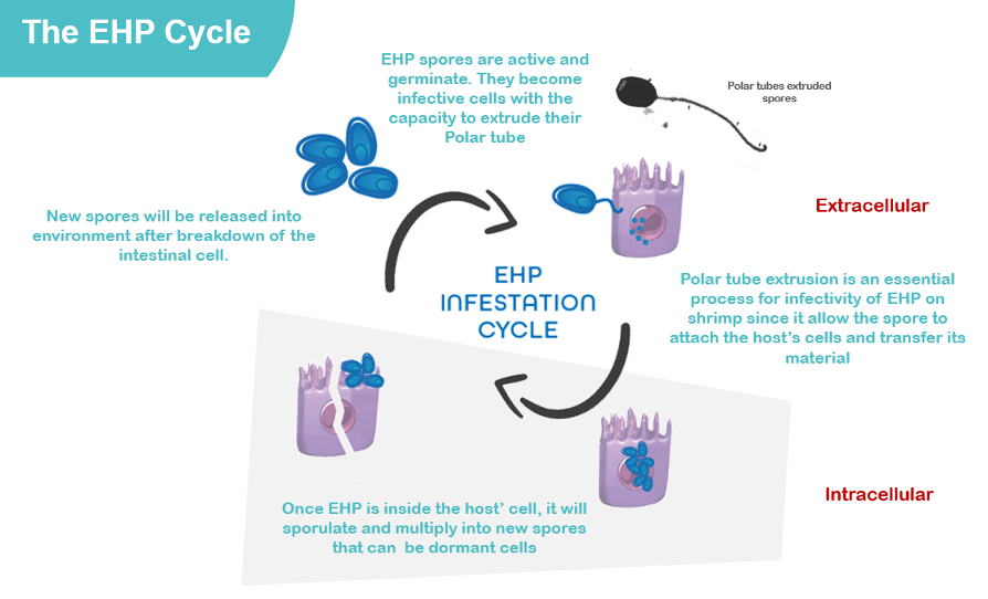
The infective phase is the only phase that can survive without the host since spores are outside of the host’s cells in a dormant and more resistant form. Once in the host, the dormant spore will become an infective one through the key phase of the cycle: the germination. This phase is notably marked by the expression of an essential tool for the spore to replicate: the polar tube.
Thanks to it, the germinated spore will puncture the plasma membrane of the host’s cell, releasing sporoplasms (infectious material) inside the cytoplasm of the cell. The sporoplasms start to replicate in many copies which are released into the environment after killing the cells, and eventually infect other shrimps.
Since EHP has an internal phase during which spores enter host’s cells, it is important to ensure that the parasite is inhibited from host cell entry during its external phase when spores are free, non-protected and before the emergence of their polar tube. This is a fundamental point when deciding on recommendations for application of this new biosecurity tool.
A specific solution designed to reduce the EHP load and its impacts
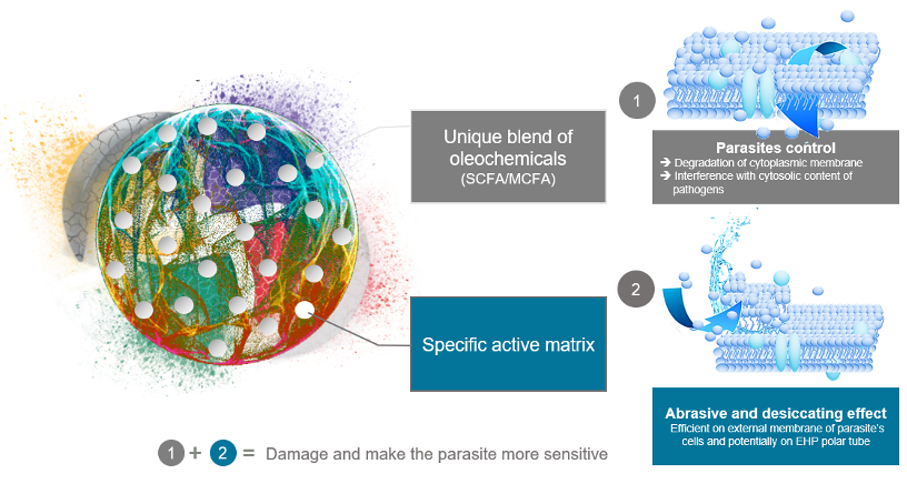
Laboratory and field trials demonstrated that a feed solution (A-Coverost, MiXscience) based on an innovative blend of oleochemicals associated to a specific active matrix (Figure 2) significantly reduces the capacity of EHP spores to germinate and express the polar tube directly linked to their capability to be infective. This effect clearly impacts the final EHP load in hepatopancreas, impacting the whole infection dynamic in the pond/farm and helps to maintain good shrimp performance to reach commercial sizes.
In vitro results
At the laboratory scale, the EHP spores used were purified according to a standard protocol developed by the center (Aldama-Cano et al., 2018). The purified spores were aliquoted and incubated with the experimental product (A-Coverost) for 30 minutes at room temperature (27°C) in Eppendorf tubes and a total volume of 1mL per reaction. The effect of the product on germination and spore viability was measured by microscopic observation after a staining step with phloxine. The germination rate or percentage of spore extrusion was calculated from the number of spores extruded (or germinated) out of the 100 spores observed.
Results showed that A-Coverost significantly reduced (>60%) the germination rate of EHP spores. It has been expressed by a lower expression of polar tubes visible under microscope. This trend has been observed in the two consecutive trials demonstrating a certain robustness of the efficacy of the product (Figure 3).

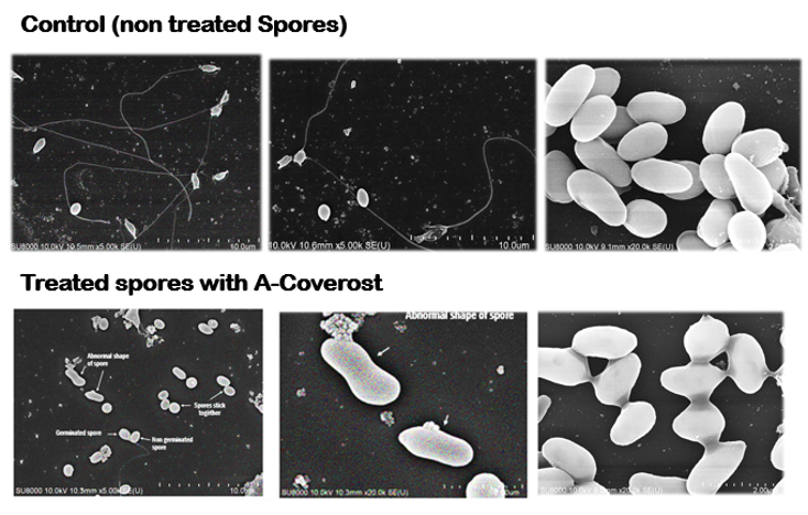
Complementary studies involving scanning electron microscopy (SEM) coupled with epifluorescence microscopy (and in particular with markers of cellular integrity and viability) demonstrated the fine mechanisms of actions of the products to explain how the it disturbs the EHP spores and their germination.
These involved the evaluation of the direct effect of A-Coverost tested at 2, 4, 6 and 8g/L on EHP purified spores. Monitoring by SEM allowed us to understand the impact of the product (tested at different dosages) on the morphology of the spores, their ability to germinate and therefore their potential in infecting shrimp. In parallel, a marking with acridine orange (AO) and propidium iodide (PI) was implemented to measure the impact on spore viability.
Acridine orange (AO) is a red/orange-fluorescent dye used in epifluorescence microscopy and flow cytometry. When AO is used, it helps to detect DNA denaturation or to detect DNA damage (Darzynkiewicz et al, 2004 and Evenson et al, 1980). Propidium iodide (PI) is a red-fluorescent dye. The use of PI is not exclusive only to live cells, it is also commonly used to detect dead cells in a population. Propidium iodide is used as a DNA stain in flow cytometry or epifluorescence to evaluate cell viability. Propidium Iodide is not membrane-permeable, making it useful to differentiate necrotic, apoptotic and healthy cells based on membrane integrity (Suzuki et al, 1997).
SEM and Epifluorescence microscopy studies
The EHP spores used were purified according to a standard protocol developed by the center (Aldama-Cano et al., 2018). The active purified spores (1×10⁷ spores) were mixed with A-Coverost at the final concentration of 8g/L. The solutions were incubated for 120 minutes at room temperature (25-26°C), before being subjected to centrifugation at 10,000rpm for 5 min.
The treated spores were washed twice with 1X PBS (phosphate-buffered saline) and 10 µL were placed on a round coverslip which were then placed in a 24-well plate and allowed to air dry. The dried coverslips were fixed with 2.5% glutaraldehyde in 0.1M buffer incubated at 4°C overnight. Treated spores on coverslips were washed twice with 0.1M PB for 10 min each and fixed with 0.1% osmium tetroxide in the dark. Spores were washed twice with 0.1 MPB for 10 min and dehydrated with alcohol in series, drying with liquid CO2, and spray coating with gold. The morphology of EHP spores was examined using a Hitachi SU8010 scanning electron microscope.
Acridine orange (AO) and PI were diluted in PBS. The two products were mixed at a ratio of 1:1 and 3μL of AO/IP mixture were mixed with approximately 20μL of spore sample. The solution was placed on a slide then analyzed under an epifluorescence microscope
The In vitro results indicated the following bullet points :
•The A-Coverost product interacted with the membrane of EHP spores, causing disruption; abnormal morphology of the spores was observed and they were no longer able to germinate (which confirmed our previous results) and tended to clump together (Figure 4).
•The A-Coverost product induced a loss of sealing of the spores, which involved the marking of the DNA with PI and therefore a reduction in the viability of the spores. We observed a very marked dose effect with an average mortality of the spores of 73% when they were exposed to a dose of A-Coverost of 4g/L and a mortality of 95 and 97% when the spores were respectively exposed with doses of 6 and 8g/L (Figure 5).
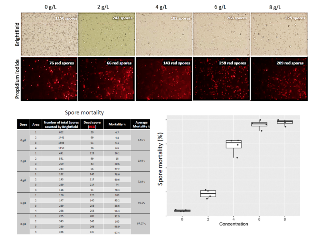
Bottom = Dose-response effect for A-Coverost concentrations ranging from 0 to 8g/L
In vivo results
Based on the previous results, the product has been tested directly in shrimp to confirm its observed potential at lab scale.
Experimental shrimp studies were performed at the BIOTEC facilities (Thailand) and at SHRIMPVET facilities (Vietnam). Following the laboratory tests, the experimental product A-Coverost was incorporated either by top coating or directly during the pelletising process. The recommended dose was 4 kg/tonne of feed for both applications.
The trials were set up as a completely randomised designs in which two groups (Challenged CTRL and Challenged A-Coverost) were compared (Table 1). Each tank contained at least 200 shrimp with an initial weight of about 3g (water volume 320L; salinity 20ppt) and shrimp were pre-fed with the indicated feed for a pre-feeding period of seven days. On day 7 of feeding, 12 shrimp from each group were collected for the detection of EHP level by qPCR. Each group had three replications.
Determination of EHP load
Shrimp hepatopancreas was collected individually from 12 shrimp per tank for DNA extraction. DNA template for qPCR was obtained by homogenising hepatopancreas tissue in DNA lysis buffer (Munkongwongsiri et al., 2021). The 20µL of qPCR reaction mixture contained 1x SYBR green-qPCR, 0.2 µM SWP primers and 10ng of DNA template. To calculate the EHP load, the samples were compared with the standard obtained from plasmid DNA at a concentration of 1.0×10⁶ copies/µL.
Parameters studied
The shrimp growth performance indicators including initial weight, final weight, weight gain, average daily growth (ADG), feed conversion ratio (FCR) and survival rate were determined.
The in vivo results indicated the following bullet points :
•The shrimp trials, carried out from 2021 to 2023, notably with EHP cohabitation approaches (EHP infection by cohabitation model (Salachan et al., 2017)) confirmed the effect of the product under different contexts and breeding conditions: country, Vietnam versus Thailand; shrimp genetics; EHP strains; food, pond conditions, etc.
•A-Coverost, applied at 4 kg/tonne of feed, has the same efficacy when applied directly by pelleting or indirectly by top coating [Figure 6].
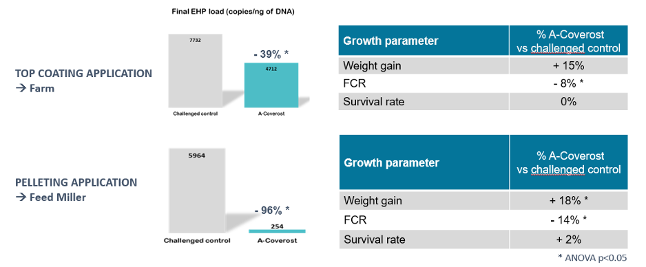
•Based on the cycle of infection of EHP, the preventive approach remains our recommendation to ensure that spores are killed or inactivated during their free phase out of the hepatopancreas cells. The sooner is the application, the better is the result. Our trials showed that it’s more pertinent to apply the product when post larvae are young.
After the pre-feeding period, shrimp from each group were transferred to three aquaria with 35 shrimp each (water volume 30L; salinity 20ppt) and placed into three cohabitation tanks (water volume 300L; salinity 20ppt) for induction of EHP infection by cohabitation model (Salachan et al., 2017).
A number of 10 EHP heavily infected shrimp, determined by a nested PCR method were added into the challenge tanks. During cohabitation, shrimp were continuously fed with the indicated feed formula. On day 17, 12 shrimp per basket were collected for the detection of EHP level by SWP-PCR. Experimental timelines are described in Table 1.
The shrimp trials confirmed the ubiquitous potential of the product to control EHP infestation in shrimp.
We understand that a direct interaction between the product and the EHP spores during their extracellular phase is a prerequisite to initiate the inhibition process and to kill the spores. This will slow down the whole dynamic of infection to control the EHP load and allow the shrimp to reach commercial sizes.. Prevention remains/is then the key approach.
However, it is clear that a single solution is not sufficient to fully eradicate EHP occurrences.
A-Coverost needs to be used as a tool in a complete management program based on biosecurity practices to implement at the farm level in order to limit the presence of the parasite.
Published in the May 2024 issue of International Aquafeed.